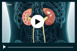
Haematuria
Haematuria is a common condition and one which must be taken seriously. Haematuria simply means blood in urine. If you notice blood in the urine it should always be investigated, although in most cases no serious cause will be found.
Haematuria is usually divided into macroscopic (where the urine is discoloured) and microscopic (where the blood is found only on dipstick or microscopy examination). Further clinically relevant distinctions can be made between painful and painless haematuria, and haematuria of glomerular and post-glomerular origin.
Haematuria investigation has been made simple with the advent of flexible cystoscopy, where the patients can assessed quickly with a local anaesthetic outpatient procedure.
Investigations for Haematuria
General Physical Examination which includes blood pressure, pulse, prostate in a male and the gynaecological organs in a female
Urinanalysis- A mid stream specimen of urine for microscopy of red, white blood cells and bacteria. The presence of any crystals, ova or parasites should be noted and culture of urine specimen. The level of protein in the urine will be assessed.
Blood tests- All patients should have a full blood count with an erythrocyte sedimentation rate. Serum urea, creatinine and electrolytes should be measured, along with albumin, calcium and liver function tests if the patient is unwell or in renal failure.
Ultrasound
CT Scan
If no abnormality is found then a flexible cystoscopy under local anaesthetic may be performed, but if either the imaging or this endoscopic examination suggest a bladder lesion the patient will require a transurethral biopsy and examination under anaesthetic for both treatment and diagnosis.
In any of the above scenarios it is important to remember that if a particular investigation pathway leads to a negative result, consideration should be given to carrying out the rest of the other pathways. Thus flexible cystoscopy for a patient with persistent microscopic haematuria in whom no renal cause is found, and ultrasound in a patient with a normal bladder and intravenous urogram.
Points to consider about Haematuria (Blood in urine)
- Haematuria may not always be a bad thing,
- Haematuria can be detected in the urine during a menstrual period.
- It can occur only during a urine infection.
- Sometimes some medicines and foods can colour the urine red. This is not the same as passing blood.
- It can occur following strenuous exercise.
Haematuria can originate from the kidney itself due to inflammation in the kidney, eg glomerulonephritis affecting the filtering units (glomeruli). When this is the cause of haematuria there are often other signs of kidney disease such as Protein in urine, High blood pressure or Abnormal renal function
Kidney cysts, tumours or kidney stones can also cause haematuria. Blockages or stones in the tube to the bladder (ureter) may cause haematuria. The bladder may also be the cause of haematuria, in cystitis (bladder infection), stones, or tumours in the bladder.
Diseases of the prostate gland may also cause haematuria
Some conditions associated with haematuria
Renal Tumours
The commonest primary renal tumour is renal cell carcinoma, an adenocarcinoma of collecting tubule origin. It commonly presents with haematuria although most are nowadays picked up incidentally by ultrasound scanning. Diagnosis is made by CT scanning and treatment is by surgical excision. Small tumours may now be treated by local excision with preservation of kidney function.
Transitional Cell carcinoma of the renal collecting system usually gives haematuria. Diagnosis may be difficult, requiring retrograde imaging and ureteroscopy. Treatment is by either local excision or, for high grade or larger lesions, nephro-ureterectomy. Immunotherapy is used for metastases with limited success; radiotherapy has little place except for palliation of bone metastases.
Benign renal tumours may cause both bleeding and diagnostic difficulty. They are, with the exception of the incidental and usually asymptomatic renal cyst, rare. Angiomyolipoma is a hamartomatous lesion, which may grow to great size and be associated with major haemorrhage; treatment is again surgical, conserving normal renal tissue where possible.
Renal Stones
Stone disease is very common, with concretions forming in the renal papillae, which then form a nidus for stone formation in the collecting system. While most stones may cause infection, one particular type (infection or matrix stone) is thought to be caused by bacteria that are able to split urea to form ammonium. Renal stones tend to be asymptomatic but may cause haematuria by either infection or direct irritation of the mucosa. They may also cause renal pain if large enough or obstructing. Diagnosis is by imaging, usually intravenous urography. Renal stones can usually be treated by extracorporeal shock wave lithotripsy on an outpatient basis, although large or complex stones may need percutaneous or open surgical removal.
Glomerulonephritis
Glomerulonephritis tends to present with microscopic haematuria. While pain may be associated, most cases will have either no symptoms or may show signs of renal failure. Investigation is as outlined above.
Pyelonephritis (ascending urinary tract infection)
Acute bacterial pyelonephritis results from bacteria ascending from the bladder either by direct spread (vesico-ureteric reflux) or possibly by periureteric lymphatic extension. Painless haematuria may occur but the symptom complex usually includes loin pain, fever and possibly septicaemia.
Papillary Necrosis
This condition occurs in diabetics and in patients with deficiencies of oxygenation, particularly sickle cell disease. It is characterised by a radiolucent filling defect on IVU and may usually be treated expectantly
Ureteric Stones
Stones may form in the kidney and drop into the tube to the bladder (the ureter ). They usually present with pain but may have haematuria as the only symptom. The presence or absence of obstruction and the size of the stone dictates management. Most ureteric stones will pass on their own but sometimes treatment by passing a telescope up to the stone to remove it is required.
Cystitis
Typically cystitis is painful and in men is commonly associated with bladder outflow obstruction. Schistosomiasis and drug related cystitis are rarer causes of bladder inflammation causing bleeding. Diagnosis is by urine microscopy and culture, assisted by cystoscopy and biopsy if necessary.
Bladder Tumours
Most of the interest in painless haematuria stems from the desire to diagnose bladder tumours at an early stage. Nearly all are transitional cell cancers, with smoking and aromatic hydrocarbon exposure being risk factors. Rarer bladder tumours include adenocarcinoma (usually arising from the urachus) and squamous cancer (associated with chronic inflammation and schistosomiasis).
Diagnosis is as outlined above with management depending on the stage and grade: 70% are superficial at presentation and are managed by transurethral surgery with or without the use of intravesical therapy. For invasive tumours the choice lies between radical cystectomy or radiotherapy. Metastatic disease may respond to platinum based chemotherapy.
Prostate Tumours
Benign prostatic hyperplasia is ubiquitous but rarely bleeds on its own: it may acute cystitis and in this case transurethral surgery is indicated. Diagnosis is by urinary flow assessment and bladder residual volume measurement. Prostate specific antigen levels should be checked to rule out prostate cancer, which while uncommon in the fifties does occur and may cause haematuria directly or by infection.
Diagnosis is by prostatic biopsy, usually with ultrasound control. Treatment depends on the stage and outlook, but local disease may be suitable for radical prostatectomy or radiotherapy while advanced disease responds to hormonal manipulation.
Rare Causes of Haematuria
Arteriovenous malformations, tuberculosis and arteritis may all cause haematuria. Patients on anticoagulants whose control is in the normal therapeutic range and who have haematuria must be fully investigated as above, since haematuria is not a normal consequence of anticoagulation

 Menu
Menu


